Have you ever wondered if a little glowing medicine could show you what’s happening inside your body? Nuclear medicine uses tiny amounts of special medicine (called radiopharmaceuticals) that light up your organs. It gives doctors a live look at how your body works, almost like watching a movie from the inside. And guess what? It doesn’t just find problems; it sometimes helps treat them too, offering real-time clues for better care. Instead of only showing your outside, this technology reveals how your insides function, helping healthcare teams make smarter, more informed decisions about your well-being.
Nuclear Medicine Fundamentals: Radiopharmaceuticals, Gamma Cameras, and Dual Diagnostic-Therapeutic Roles
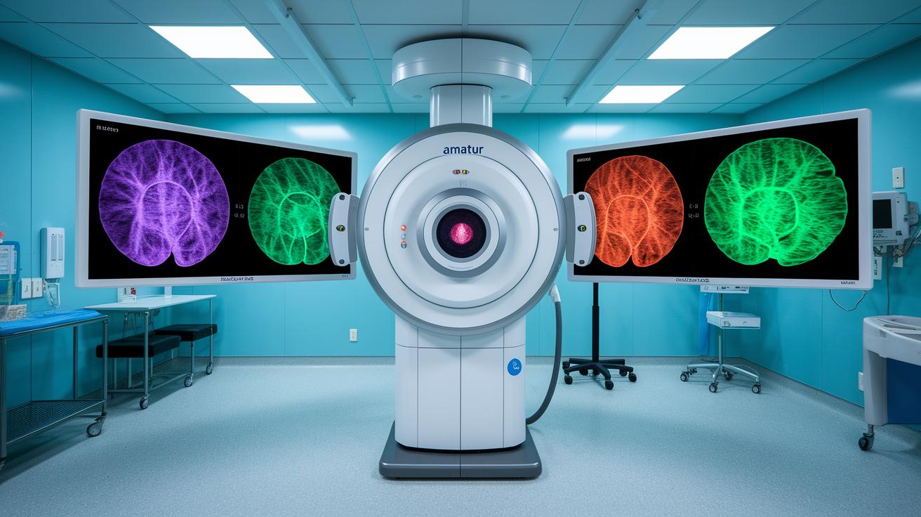
Nuclear medicine uses tiny doses of a radioactive medication called radiopharmaceuticals. These are injected into the bloodstream so that organs briefly “light up,” making them easier to study. In this way, a gamma camera can capture the short burst of radiation as it leaves the tissues, showing us real-time images of how your organs are functioning. Imagine that for a moment, you actually become the source of light, and your body tells its own story from the inside.
This method differs from traditional X-rays or CT scans that merely cast shadows. Nuclear imaging shows how organs work rather than just how they look. It’s like watching a live performance in your body, full of dynamic details.
On top of that, advances in radiopharmaceuticals now allow the same substances used for diagnosis to also treat diseases. Take iodine-131, for example. This substance helps treat thyroid disorders by zeroing in on thyroid cells while also highlighting areas that may need extra attention. By combining both diagnostic and treatment roles, doctors can better understand what's happening inside and make more informed decisions for your care.
Advanced Nuclear Medicine Techniques: PET, SPECT, and Hybrid Imaging Modalities
PET scans use a special tracer known as FDG to show how active your cells are by lighting them up almost like tiny bulbs. It gives us a quick peek into how energy moves through your body, often spotting early changes before they show up on a regular scan.
SPECT works in a similar way but uses tracers that send out gamma rays. With the help of gamma cameras, it helps experts spot problems that normal tests might miss. For example, if a SPECT scan shows uneven blood flow in your heart, it might signal a condition that needs more attention.
Hybrid imaging takes things a step further. Systems like PET/CT and SPECT/CT combine the metabolic snapshots from the tracers with clear anatomical pictures. This means doctors can see not only how your tissues work, but also the exact spot where something might be wrong. Sometimes, PET/MRI is chosen to get a better view of soft tissues, which is especially useful for checking areas like the brain or pelvis.
These procedures usually take between 30 and 60 minutes. However, some tests, such as bone scans, might need a few hours to allow enough tracer to accumulate. By tracking how these radiotracers move in your body, clinicians can pick up on subtle changes that might be an early sign of illness.
Molecular imaging is always pushing forward, making sure each scan delivers key insights to help guide your care.
Clinical Applications of Nuclear Medicine: From Oncology to Cardiac and Endocrine Evaluations
Nuclear medicine helps doctors spot and treat many health issues like cancer, thyroid problems, heart disease, and liver troubles. FDG-PET scans are a big part of cancer care, they catch tumors, show how advanced they are, and check how well treatment is working. For instance, a PET scan can pick up tiny changes in cancer cells, even giving clues about certain blood cancers. Radioactive iodine, known as I-131, is used to both diagnose and treat thyroid problems. It works by measuring how much iodine the thyroid absorbs and then giving a targeted treatment right where it's needed.
Bone scans use special technetium-based tracers to check bone density and find any spread of cancer. Heart doctors use myocardial perfusion imaging to see how well blood is flowing through the heart, which is key for diagnosing coronary artery disease. In addition, hepatobiliary scans give important clues about how well the liver is working and how bile moves through the body.
For children, the medicine doses change based on weight to keep things safe and effective. New treatments using targeted radioisotopes are focusing directly on tumors. For more on this cutting-edge approach, check out this link on Personalized Medicine.
| Application | Radiotracer | Technique | Clinical Use |
|---|---|---|---|
| Oncology | FDG | PET scan | Cancer detection, staging, monitoring |
| Endocrine | I-131 | Whole-body scan | Thyroid uptake measurement, therapy |
| Bone Evaluation | Technetium-99m agents | Bone scintigraphy | Checking bone density, spotting metastases |
| Cardiac | Technetium-99m sestamibi | Myocardial perfusion imaging | Measuring blood flow in the heart |
| Hepatic | HIDA agents | Hepatobiliary scan | Assessing liver function and bile flow |
Radiation Safety and Dosimetry in Nuclear Medicine Practice
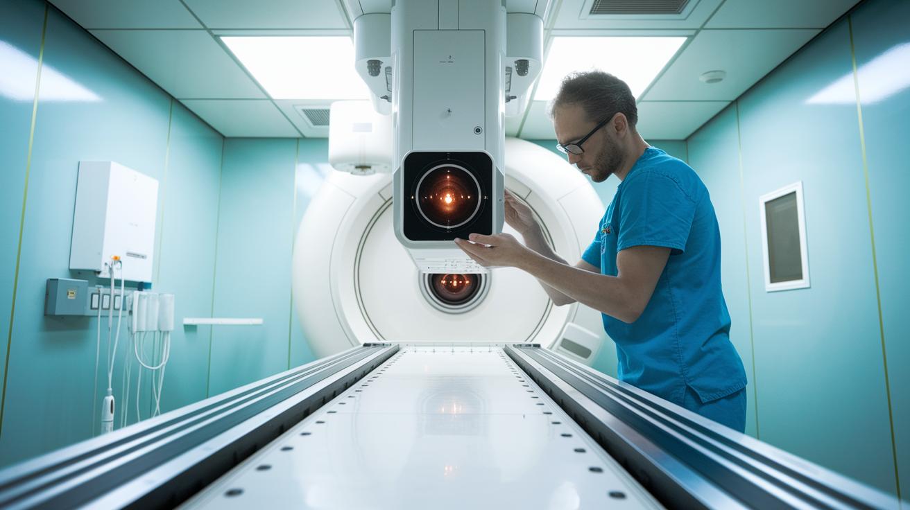
Nuclear medicine uses very low doses of radiation to keep both patients and staff safe. Every step is taken to ensure that only the tiny, necessary amount of radiation is used for clear, detailed images, imagine using just the right amount of paint to reveal a hidden masterpiece.
To get that perfect dose, professionals carefully measure the activity of radiotracers and adjust the gamma cameras as needed. This process is backed up by regular equipment checks and tests to make sure the radiotracers are pure. Think of it like a musician tuning their instrument right before a big performance.
Patient safety is at the core of these procedures. Customized dosimetry calculations make sure that you get the most useful images with the least possible risk, even taking rare allergic reactions into account with extra precautions. After your scan, you get clear instructions on how to handle the brief period of residual radioactivity, so caregivers and family members stay safe too. With consistent quality checks and careful monitoring, every phase of the process ensures both reliable imaging and strong radiation protection.
Nuclear Medicine Empowers Bold Clinical Care
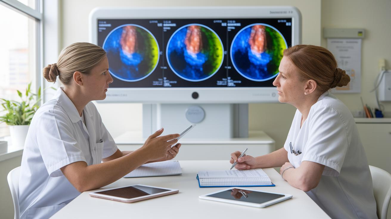
Imagine having a smart buddy in the clinic , that's what artificial intelligence does for nuclear imaging. It helps doctors get a crisper, faster look at scans by using smart algorithms to fill in details. Think of it like noticing the first glow of dawn when you wake up. Researchers are also speeding up studies on new tracers to catch tiny signs of trouble before they become big issues. It’s a bit like spotting an early clue in a mystery novel.
Hybrid imaging systems like PET/MRI and SPECT/CT now mix functional details with clear anatomical snaps. This means doctors can see exactly where a problem is hiding. New designs for how scanners capture images, plus the ability to share scans via the cloud, let experts consult with each other from anywhere. These updates offer clear, quick insights that empower doctors to take confident, bold steps in treatment.
Regulatory changes are also smoothing the way for new radiopharmaceuticals. In short, this wave of tech improvements and research breakthroughs is giving clinical teams the tools they need to deliver more focused, responsive care in nuclear medicine.
Professional Roles in Nuclear Medicine: Training, Certification, and Interdisciplinary Collaboration
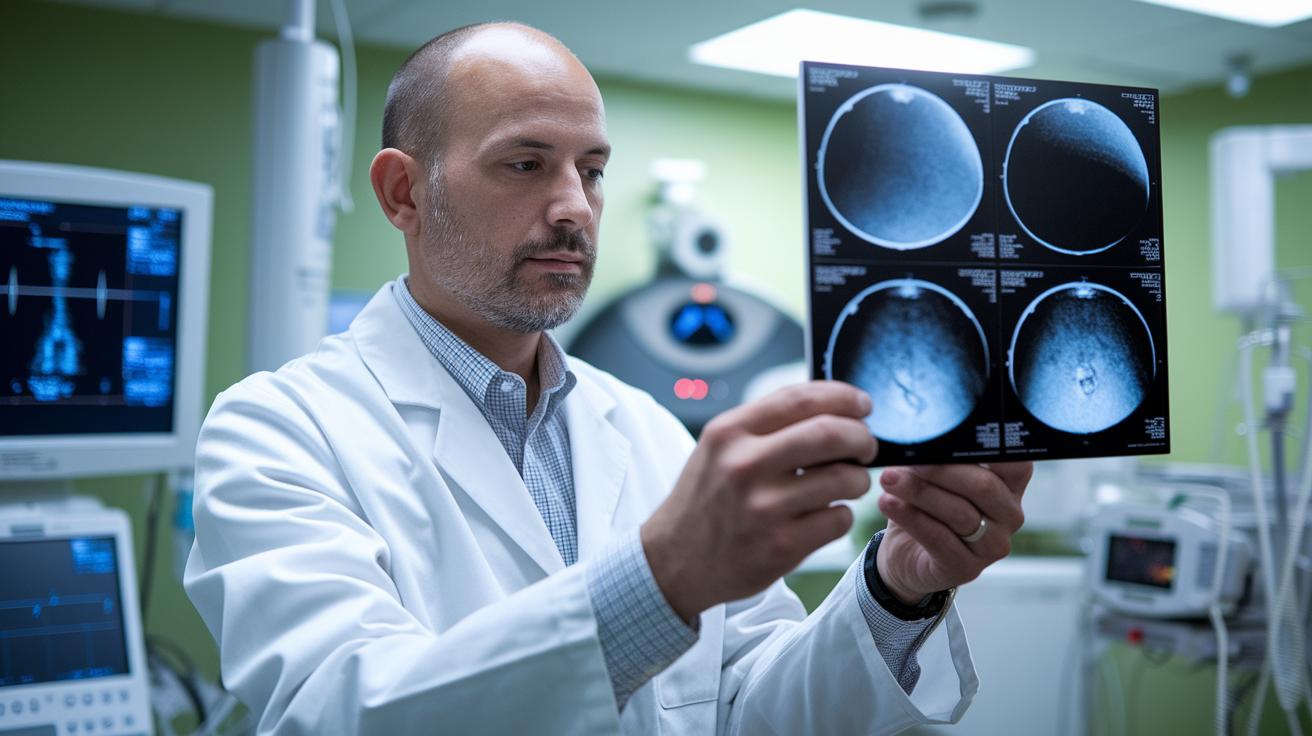
Imagine a nuclear medicine doctor who’s spent years in a specialized residency program that mixes studies in radiology, cardiology, or oncology. After long hours of learning and hands-on practice, they become board-certified experts ready to dive into clinics and interpret complex scans with clarity and confidence.
Technologists in this field follow a similar path of dedication. They earn university degrees and get certified in handling radiopharmaceuticals (special medicines used in imaging) and operating advanced equipment. Their education covers everything from keeping gamma cameras in top shape to fine-tuning dosing protocols. And because health technology is always evolving, they stay on their toes with continuous learning and regular accreditation updates.
Teamwork really drives nuclear medicine forward. Doctors, technologists, and specialists like oncologists, cardiologists, and medical physicists work closely together to crack diagnostic challenges. This collaborative spirit means every expert shares their unique insights, leading to better patient care and more balanced clinical outcomes.
This team-based approach not only enhances patient care but also opens up opportunities for ongoing career growth and regular reviews of educational programs.
Final Words
In the action, the article illustrated practical aspects of nuclear medicine, from radiopharmaceutical basics to sophisticated PET and SPECT techniques. Safety measures and dosimetry ensure patients and staff are properly protected. Clinical applications and emerging innovations highlight how nuclear medicine transforms diagnostics into effective, real-world care. Professional training and technological advances work together to create a more confident health landscape. Embracing these research-backed approaches inspires optimism and a brighter, healthier future.
FAQ
Nuclear Medicine near me
Finding nuclear medicine near you means locating local centers where professionals use small amounts of radioactive medication to capture images and treat conditions safely.
Nuclear Medicine doctor
A nuclear medicine doctor specializes in using radiopharmaceuticals for imaging and therapy, collaborating with other experts to diagnose health issues through functional imaging techniques.
Nuclear Medicine salary
The nuclear medicine salary varies by location, experience, and certification, often reflecting competitive pay in the medical imaging field where advanced diagnostic skills are essential.
Nuclear Medicine degree
A nuclear medicine degree involves focused study on radiopharmaceuticals, imaging technologies, and safety measures, preparing graduates for clinical roles and research in functional imaging.
Examples of nuclear medicine
Examples of nuclear medicine include PET scans, SPECT scans, and bone scintigraphy, each using tracers like FDG or I-131 to visualize organ function and detect disease early.
Nuclear medicine Radiology
Nuclear medicine radiology differs from traditional radiology by emphasizing functional imaging through radioactive tracers, which reveal organ activity rather than just anatomical structures.
Side effects of nuclear medicine
The side effects of nuclear medicine are rare and usually mild, with some patients experiencing minor allergic reactions or slight discomfort at the injection site due to low radiation doses.
Nuclear medicine meaning
Nuclear medicine means using tiny amounts of radioactive material to capture images of physiological processes, helping doctors evaluate organ function and guide targeted therapies.
What is nuclear medicine used for?
Nuclear medicine is used for diagnosing and monitoring diseases by visualizing organ function through radiopharmaceuticals and gamma camera technology, aiding in targeted treatment decisions.
What does a doctor in nuclear medicine do?
A doctor in nuclear medicine interprets diagnostic scans produced by radiotracers, collaborates with specialists, and helps manage treatments based on detailed functional imaging data.
What is the difference between radiology and nuclear medicine?
The difference between radiology and nuclear medicine is that radiology focuses on structural imaging using X-rays, while nuclear medicine highlights functional activity through radioactive tracers.
What is the highest paid nuclear medicine?
The highest paid nuclear medicine professionals are typically physicians with advanced certifications and specialized expertise, reflecting leadership roles in high-demand imaging and therapeutic services.