Ever wondered if a tiny bit of tissue could unlock big secrets for your health? Today, labs use clear digital screens and smart AI to spot details that old microscopes often missed.
Long, boring exams are now a thing of the past. Modern techniques mix simple lab tests with high-resolution imaging to deliver faster, more exact results.
This fresh approach helps doctors pinpoint problems quickly so you get the care you need sooner. Keep reading to learn how these breakthroughs are completely changing the world of pathology.
Pathology Innovations: Digital and Molecular Advancements
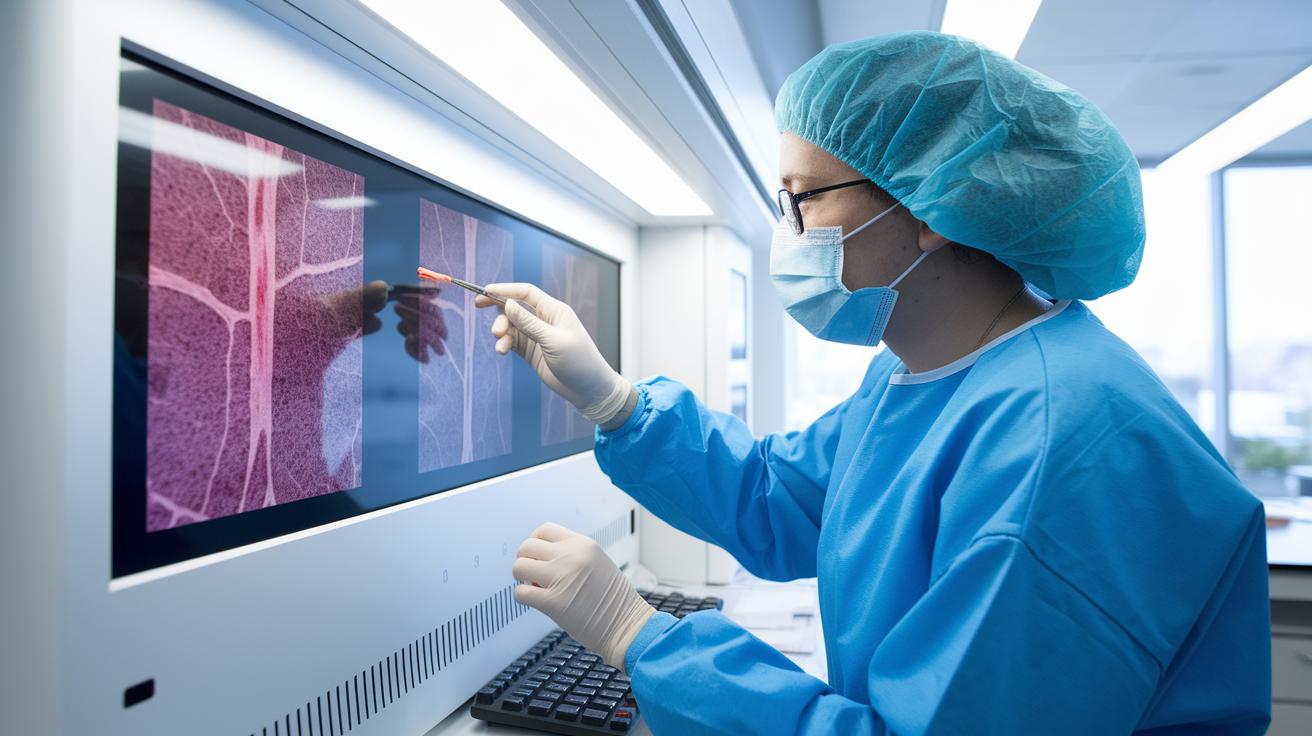
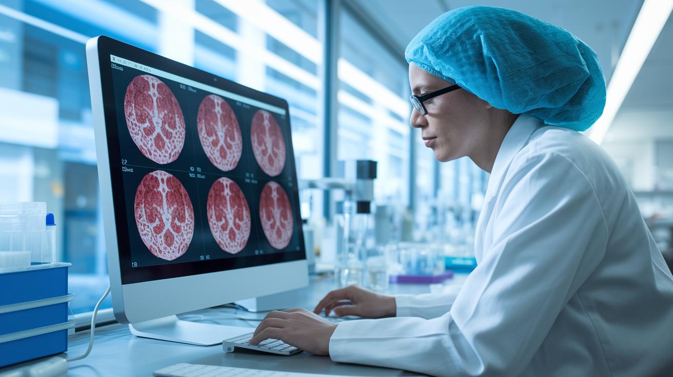
In the past, pathology meant spending hours looking at hand-prepared slides through optical microscopes. Today, we’re embracing a blend of digital tools, molecular tests, and AI to transform how tissue samples are examined. Imagine checking a tissue sample on a crisp, high-resolution screen instead of peering through a microscope, it’s a real game changer that brings clarity and speed.
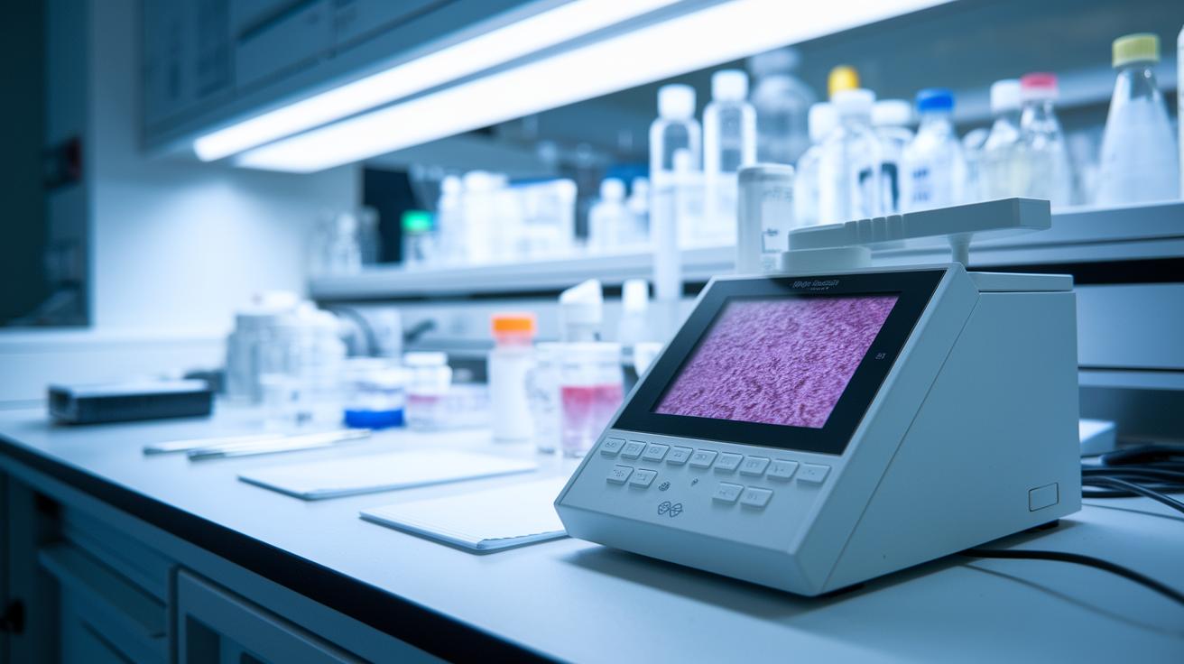
Advances like cutting-edge molecular diagnostics, smart AI algorithms, and high-quality imaging are at the heart of this transformation. With modern techniques, labs can spot tiny cellular markers that used to be missed. For example, a digital slide scanner (installed on September 9, 2024, by a North American academy) can capture images with stunning detail. These images are then analyzed by AI programs that flag any abnormalities and offer insights into what’s happening inside the cells. Think about it: a lab can now review hundreds of slides in the time it used to take to look at just a few, marking a huge leap in efficiency.
All of these advancements help make diagnoses much more precise. With digital imaging and automated tests, errors shrink and results come back faster. This means that doctors get more accurate findings quickly, helping them to outline targeted treatments sooner. In short, combining digital and molecular methods is reshaping the way research, diagnostics, and treatments work, leading to better outcomes for patients.
Digital Pathology and Imaging Diagnostics
Today, pathology isn’t just about old-fashioned microscopes. Labs are switching to digital methods that let them examine tissue samples with amazing clarity using high-resolution digital slide scanners and telepathology systems. These tools also make it easy for experts to consult with each other in real time, even from far away.
You’ll find labs using:
- High-resolution digital slide scanners
- Virtual microscopy platforms for web-based review
- AI-driven image-analysis software (where AI means computers learning to spot details)
- Telepathology systems for expert consultations
- Cloud-hosted case-management repositories
Remote diagnostic systems are a real game-changer. By merging secure cloud storage with expert reviews, they help bridge gaps no matter the distance. And thanks to digital teaching modules like USCAP’s eLearning platforms, clinicians can learn about the latest computational pathology tools and even how to spot subtle changes in tissue structure.
Molecular Diagnostics and Genomic Profiling in Pathology

Molecular diagnostics has totally changed how we spot diseases by zeroing in on small clues hidden in our genes, proteins, and cell chemicals. Labs now use smart techniques that reveal little markers that older tests often missed. For example, checking levels of biomarkers like Pancreatic Stone Protein (a marker that can indicate early sepsis) can help doctors catch sepsis before it gets serious. This means diagnoses are more accurate and treatments can be fine-tuned to each patient’s unique condition.
Next-generation sequencing and proteomic profiling take these advances even further. The former is all about reading the detailed map of your genes, picking up tiny mutations, while the latter gives a clear picture of how proteins behave and interact inside cells. These methods dig deep into the tumor’s surroundings, offering clues that help doctors decide on targeted treatments. In short, by combining these brilliant techniques, modern labs can understand even the trickiest diseases much better.
New waves in biomarker discovery and metabolomic research add another layer of precision. Metabolomic research watches how chemical changes in cells occur, helping predict how patients might respond to treatment and refining outcome measurements. When you mix fresh insights from genomic profiling with detailed protein analysis, you start to see clear patterns in how diseases develop. This complete picture, blending traditional molecular diagnostics with high-tech sequencing and protein exploration, is paving the way for precision medicine, where treatments are as unique as the patients themselves.
Artificial Intelligence Integration in Pathology Workflows
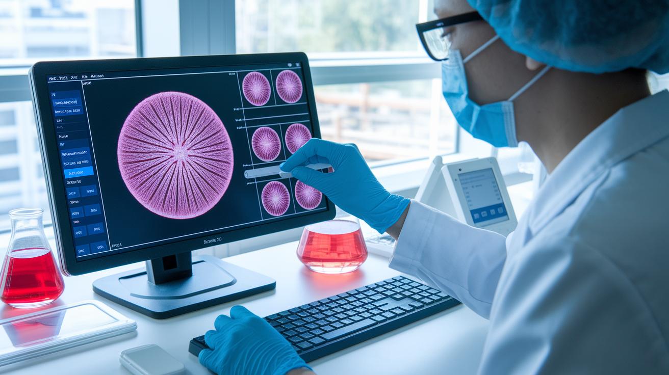
AI image analysis and smart algorithms are changing the way labs look at tissue samples. These clever tools help spot tiny details in digital slides, cutting down the time needed for manual checks by up to 30%. They learn from thousands of images, getting better and better at noticing important clues that might even slip past experienced eyes. Imagine a tool that quietly scans hundreds of images and highlights spots that need a closer look by a person, this is already happening in many labs. In short, these precise diagnostic tools help labs find unusual cells or patterns faster, which means quicker and more accurate results.
In everyday hospital settings, AI modules have become a natural part of the hospital information systems. They support quick decision-making in busy areas like the ICU, heart care, and cancer treatment. These systems also help with automatic reporting, making results more consistent and reducing mistakes. Thanks to clinical workflow automation that offers real-time insights, hospitals run more smoothly and use their resources wisely, ultimately leading to better care for patients.
Laboratory Automation and Quality Control in Pathology
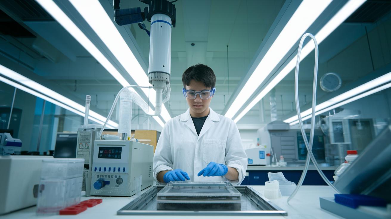
Laboratory automation is totally changing how pathology labs handle samples. Automated processing systems are now a regular part of everyday workflows. With tools like automated tissue processors and slide stainers, labs can work on large batches of slides at once, cutting down on manual mistakes and speeding up processing times.
Core automated instruments really simplify everyday tasks. For example, modern tissue processors use precise, pre-set protocols to move tissue samples through several preparation steps automatically. And slide stainers make sure every slide gets the same treatment, which is key for an accurate diagnosis. This means technicians can spend more time tackling the tougher diagnostic challenges.
Quality control is the heartbeat of these automation systems. Labs put in place strong quality assurance programs with regular checks, audits, and validation routines to ensure every step meets established standards. It’s all about linking each part of the sample handling process to real clinical outcomes, so quality is never compromised.
Best practices in lab management hinge on solid regulatory compliance and rigorous quality checks. By following proven standard procedures and performing consistent audits, labs can balance the efficiency of automation with top-notch analytical precision. Taking the extra time to validate automated systems builds reliability and trust, ultimately leading to better patient care with more consistent diagnostic results.
Case Studies and Clinical Integration of Modern Pathology Techniques
Case studies offer a clear look at how today's pathology is changing disease detection and treatment. They let us see how new methods help doctors make faster, better decisions. One study showed that the Pancreatic Stone Protein biomarker cut sepsis detection time by 12 hours. This faster diagnosis can save lives. Another case focused on a digital learning approach from the USCAP GI tract pathology tutorial, where digital tools improved hands-on training. Plus, using digital pathology to examine rare tumor settings reduced analysis time by 25 percent. These examples show how blending technology with everyday clinical work makes a real difference.
| Case Study | Technique | Key Outcome |
|---|---|---|
| Pancreatic Stone Protein Biomarker Study | Molecular diagnostics | Improved sepsis detection lead-time by 12 hours |
| USCAP GI Tract Pathology Tutorial | Digital case-based learning model | Enhanced diagnostic training |
| Rare Tumor Microenvironment Profiling | Digital pathology | Reduced analysis time by 25% |
These studies remind us that lab research is now a part of everyday care. Technology helps deliver practical insights right when they are needed. For instance, insights shared during the 2025 Medical Student Genomics Workshop showed how hands-on research informs better treatment plans. Such real-world examples build trust in new diagnostic methods and support more personalized, patient-centered care.
Future Directions and Emerging Trends in Pathology Research

New education and training programs are opening exciting pathways in pathology. For example, the 2025 Medical Student Genomics Workshop and the GI tract pathology tutorial use interactive, digital methods that transform the old classroom setup. Picture a lab where cutting-edge theory meets hands-on practice.
Mentorship academies and ambassador programs are also reshaping how we learn medicine. They pair up rising talents with seasoned experts, letting practical skills grow alongside solid theory. Whether you’re a student or a pro in the field, these dynamic training paths open up fresh opportunities.
We’re also seeing more research that blends disciplines like neurology, heart studies, and autoimmune pathology. Small teams are joining forces to tackle shared challenges, which helps us better understand complex diseases. In short, bringing different specialties together sparks innovative ideas and creative solutions.
New funding opportunities are fueling these trends, too. Fresh grant programs and targeted support are paving the way for studies that cross traditional boundaries, turning scientific breakthroughs into meaningful, real-world benefits. Expect even more collaboration and well-funded research in the future of pathology.
Final Words
In the action of blending digital tools, molecular insights, and AI-powered workflows, we see how modern pathology transforms traditional practices into dynamic, research-based routines. The article highlights how lab automation and case studies inform clinical integration, setting a clear path toward improved patient outcomes. Each section shows practical ways technology and research advance our understanding while making health decisions more informed. The future looks bright as these innovative practices empower better, balanced care for all.
FAQ
Modern pathology Impact Factor
The modern pathology impact factor indicates how frequently articles in the journal are cited. For example, a 2023 impact factor of 7.1 highlights its strong influence within the pathology community.
Modern Pathology submission
The modern pathology submission process requires authors to follow specific guidelines. This process ensures that submitted research meets quality standards and formatting requirements before it undergoes peer review.
Modern Pathology publication fee
The modern pathology publication fee covers the costs associated with editorial processing and production. Detailed fee information is available on the journal’s website to help authors plan their submission budgets.
Modern Pathology author guidelines
The modern pathology author guidelines provide clear instructions on manuscript formatting, ethical practices, and reference styles. These guidelines help authors ensure their work fits the journal’s standards for scientific rigor and clarity.
Modern Pathology editorial board
The modern pathology editorial board comprises experts in various pathology fields. They oversee the review process, helping to maintain high-quality research and strong ethical standards within the journal.
Modern Pathology impact factor 2025
The modern pathology impact factor for 2025 is anticipated to reflect ongoing research excellence and citation trends. Projections consider current research influence and emerging trends in scholarly output.
Modern Pathology current Issue
The modern pathology current issue features the latest research on digital imaging, molecular diagnostics, and innovative pathology techniques. It presents findings that advance our understanding of disease processes and diagnostic methods.
Modern Pathology case report
The modern pathology case report outlines unique clinical cases that provide valuable insights for medical diagnosis. Such reports help clinicians recognize rare conditions and improve diagnostic strategies.
Is the father of modern pathology?
The father of modern pathology is often considered to be Rudolf Virchow. His groundbreaking work in cellular pathology laid the foundation for understanding the mechanisms of disease.
What are the 4 types of pathology?
The four types of pathology generally include clinical, anatomic, forensic, and molecular pathology. Each type focuses on different aspects of disease study, from laboratory testing to examination of tissues.
What is the aim and scope of modern pathology?
The aim and scope of modern pathology center on merging traditional histology with advanced digital and molecular techniques. This integration drives innovative research and improves precision in diagnostic practices.
What are the three types of pathology?
The three types of pathology commonly discussed are clinical, anatomic, and forensic pathology. These fields cover a broad range of diagnostic methods and play crucial roles in understanding and investigating diseases.